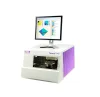
Hệ thống kính hiển vi siêu quang phổ


Tổng quan về sản phẩm Hệ thống kính hiển vi siêu phổ
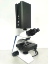
Hệ thống kính hiển vi siêu phổ là công nghệ cải tiến kết hợp các thiết bị siêu phổ và kính hiển vi, với chức năng của cả công nghệ siêu phổ và kính hiển vi. Hệ thống có thể thu được thông tin quang phổ có độ phân giải cao của các mẫu ở các dải bước sóng khác nhau, cũng như thu được hình ảnh hình thái của các mẫu sinh học, đạt được sự kết hợp giữa quang phổ và hình ảnh.
Kính hiển vi siêu phổ là công nghệ cải tiến kết hợp máy ảnh siêu phổ với kính hiển vi, cung cấp các chức năng của cả công nghệ siêu phổ và kính hiển vi. Hệ thống cho phép thu thập thông tin quang phổ có độ phân giải cao từ các mẫu ở các dải bước sóng khác nhau, đồng thời chụp được cấu trúc hình thái của các mẫu sinh học.
Sự tích hợp giữa hình ảnh và quang phổ này đạt được sự kết hợp liền mạch giữa các phép đo và quang phổ, cung cấp phân tích quang phổ chính xác hơn. Sự tiến bộ đột phá này trong công nghệ tích hợp các lĩnh vực phép đo và quang phổ thông qua hình ảnh siêu phổ, cho phép thu thập đồng thời thông tin không gian và quang phổ.
Những ưu điểm chính của hệ thống bao gồm: Độ chính xác cao: Độ phân giải quang phổ ở cấp độ nanomet với phạm vi bước sóng bao phủ khắp các vùng UV, khả kiến và hồng ngoại. Hiệu suất cao: Phát hiện tốc độ cao và xử lý dữ liệu nhanh chóng.
Khả năng tại chỗ: Không cần chia sẻ hoặc di chuyển đối tượng được đo. Không phá hủy: Chỉ cần chiếu sáng cần thiết mà không cần tiếp xúc vật lý hoặc làm hỏng mẫu
Hướng ứng dụng của Hệ thống kính hiển vi siêu phổ

Sinh học tế bào
1.Cấu trúc vi mô của tế bào (cơ quan, ty thể, v.v.)
2.Chụp ảnh tế bào sống
3. Các quá trình sinh học của tế bào
• Sinh học nano: Tương tác giữa các hạt nano và tế bào
• Nghiên cứu về cơ quan và thuốc
• Nghiên cứu về bề mặt và cấu trúc vật liệu
• Bệnh lý học và cấu trúc cắt ánh sáng
• Nghiên cứu thực vật học (bao gồm thuốc lá)


Các trường hợp ứng dụng của kính hiển vi siêu phổ
Kính hiển vi siêu phổ có thể được ứng dụng trong nhiều lĩnh vực nghiên cứu sinh học, đặc biệt là trong mô bệnh học, sinh học tế bào và vật liệu sinh học. Chúng cung cấp hình ảnh huỳnh quang có độ phân giải cao kết hợp với phân tích quang phổ theo vị trí cụ thể, cho phép nghiên cứu chi tiết ở cấp độ tế bào và cấu trúc nano.

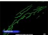
Mẫu mô
Hình ảnh hình thái mô học và đường cong quang phổ tương ứng cho từng phần

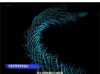
Mẫu sinh học tế bào
Hình ảnh hình thái tế bào học và đường cong siêu quang phổ tương ứng

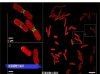
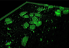
Mẫu sinh học đơn bào
Đường cong quang phổ tương ứng với từng phần của tế bào

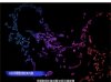
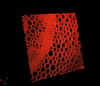
Mẫu vật liệu sinh học nano
Đường cong quang phổ tương ứng với từng phần

Hạt nano trong tế bào ung thư~ Đường cong hình thái và quang phổ tương ứng

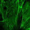
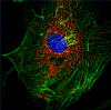


Example: High-resolution fluorescent images

Hyperspectral ultra microscopic system~ 3D image construction
Ứng dụng với Kỹ thuật huỳnh quang
Việc sử dụng các kỹ thuật huỳnh quang (Ứng dụng huỳnh quang) trong chẩn đoán y khoa trở nên mạnh mẽ hơn khi được tích hợp với các hệ thống kính hiển vi siêu phổ. Kính hiển vi siêu phổ là công cụ có khả năng phân tích tế bào ở cấp độ vi mô và dưới tế bào với độ chính xác và độ phân giải đáng kinh ngạc. Chúng đóng vai trò quan trọng trong các lĩnh vực như chẩn đoán ung thư và phát hiện bệnh truyền nhiễm bằng cách cung cấp kết quả chính xác và cục bộ.
Lợi ích của Ứng dụng này
- Phát hiện đồng thời nhiều chất huỳnh quang (Đa kênh)Kính hiển vi truyền thống thường không thể phân biệt các chất huỳnh quang có phổ phát xạ tương tự (ví dụ: xanh lục-vàng). Ngược lại, kính hiển vi siêu phổ có thể tách các tín hiệu này thành các phổ chi tiết, cho phép người dùng xác định các nguồn riêng biệt ngay cả khi có bước sóng chồng lấn.
- Độ chính xác cao hơn với chức năng ngăn chặn tự huỳnh quang
Các mẫu sinh học thường biểu hiện tự huỳnh quang (huỳnh quang tự nhiên từ tế bào), có thể gây trở ngại cho việc phát hiện. Hệ thống siêu quang phổ có thể phân biệt giữa tín hiệu mục tiêu và huỳnh quang tự động nền hiệu quả hơn so với các hệ thống thông thường. - Chụp ảnh quang phổ trong tế bàoKỹ thuật này cho phép các nhà nghiên cứu không chỉ nhìn thấy vị trí huỳnh quang mà còn xác định hóa chất hoặc protein nào có mặt tại mỗi vị trí, cho phép phân tích phân tử chuyên sâu
- Phân tích không phá hủy
Cho phép chụp ảnh tế bào sống bằng cách sử dụng huỳnh quang để theo dõi hành vi tế bào và các sự kiện sinh hóa theo thời gian thực, mà không làm hỏng mẫu.


Loại nghiên cứu nâng cao:
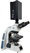
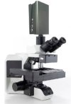
Hệ thống kính hiển vi siêu phổ
Hệ thống kính hiển vi siêu phổ mới này có thể dễ dàng thu được hình ảnh huỳnh quang có độ phân giải cao, giúp các nhà nghiên cứu thu được các cấu trúc vi mô mẫu tốt hơn.
Hệ thống này có thể được sử dụng rộng rãi trong các lĩnh vực như sinh học tế bào,
khoa học vật liệu, dược phẩm, huyết học và miễn dịch học y khoa.
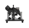
Loại nghiên cứu : Hệ thống kính hiển vi siêu phổ
(Dòng huỳnh quang thẳng đứng và đảo ngược)
Các Sản Phẩm Liên Quan
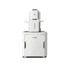
Hyperspectral Microscope Specifications [Mock up]

Dịch vụ Khách hàng


