What is an Electron Microscope, What are Its Advantages, and How Many Types Are There?

What is an Electron Microscope?
An electron microscope is a scientific tool that uses a beam of electrons instead of light to create high-resolution images of tiny objects. Because electron beams have much shorter wavelengths than visible light, they can achieve far greater magnification much higher than traditional light microscopes.
Types of Electron Microscopes
There are 3 main types of electron microscopes:
1. Transmission Electron Microscope (TEM)
TEM uses a high-energy electron beam to pass through an ultra-thin sample (about 50100 nanometers). It provides 2D high-resolution images, capable of showing atomic-level structures. It's widely used in materials science, cell biology, and virology.
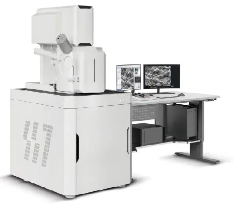
2. Scanning Electron Microscope (SEM)
SEM scans a focused electron beam across the surface of the sample. It produces 3D-like surface images with depth and high detail. It can magnify up to 12 million times, with resolution down to 120 nm. SEM is popular in material characterization and biological sample analysis.
3. Field Emission Scanning Electron Microscope (FESEM)
FESEM is an advanced version of SEM, using a field emission source to generate a stable, high-brightness electron beam. It provides superior resolution (up to 0.51 nm) and operates at low voltages ideal for non-conductive and delicate samples.
Advantages of Electron Microscopes
- Much higher magnification and resolution
- Capable of imaging atomic-level structures
- TEM produces 2D images, SEM produces 3D surface images
- Advanced models include chemical composition analysis via EDS or WDS
- Greater depth of field than optical microscopes
- Quantitative analysis possible, e.g. particle size, layer thickness, porosity
Using Electron Microscopy to Image Cancer Cells
Electron microscopes can visualize cancer cells in extremely high detail, offering benefits such as:
- Showing structures that are invisible under light microscopes
- Differentiating cancer cells from healthy ones based on organelle structure (nucleus, mitochondria, membrane)
- Tracking the spread of cancer cells in tissues precisely
How Do Electron Microscopes Work?
The operation consists of several key steps:
- Electron beam generation using a tungsten filament or field emission source
- Focusing the beam with condenser and objective lenses
- Electron-sample interaction (e.g., transmitted in TEM or scattered in SEM)
- Image detection and formation (2D for TEM, 3D for SEM)
What Are Electron Microscopes Used to Study?
Electron microscopes are used to study very thin samples in areas such as:
- Materials science
- Nanotechnology research
- Cell biology
- Medical science
- Pharmaceutical R&D
- Ceramic industry
- Semiconductor inspection
- Geology
- Food and agricultural research
Hong Kong NTI FESEM Solutions Provider
At Hong Kong NTI, we provide and distribute high-performance FESEM systems from industry-leading brands. With over 15 years of global experience, we support micro- and nano-level research and development with advanced measurement tools and expert consultation.
Visit us at 725 S-Metro Building, 20th Floor, Sukhumvit Road, Bangkok
Tel: 02-821-5278
Email: info@hknti.com
Line: https://line.me/R/ti/p/@816txpya


